Radiation therapy (radiotherapy) uses high energy x-ray beams to disrupt the DNA in cancer cells, to kill or control their growth.
It’s a localised treatment affecting only the area which is specifically targeted. Although some healthy tissue may be in the treatment area, it generally has the ability to repair itself, unlike cancer cells.
In early breast cancer, radiation therapy is used with the aim of getting rid of any malignant or pre-cancerous cells remaining in the breast following partial mastectomy or lumpectomy. This reduces the risk of developing a local recurrence of cancer in the breast in the future. Radiation therapy is also used to treat the chest wall after mastectomy if the cancer has high-risk features.
The regional lymph nodes in the axilla (underarm), supraclavicular fossa (above the collarbone) or internal mammary chain (close to the breast bone) may also be treated in some cases.
In these settings, large international trials have demonstrated that radiation therapy reduces the incidence of local breast cancer recurrence.
Radiation therapy is usually given after surgery, once the wounds have healed. For people needing chemotherapy, radiation is given after that treatment has been completed.
Radiation treatment is still offered as standard of care for all women post-lumpectomy in New Zealand but a clinical trial (EXPERT trial) is currently recruiting women who are at low risk of local recurrence (based on the PAM50 score of their tumour) and who are taking hormone therapy, to investigate whether adjuvant radiation could be safely omitted in this select group. The trial is recruiting at Waikato, Palmerston North, Wellington and Christchurch hospitals and is still open so the results will be many years away.
External beam radiation therapy
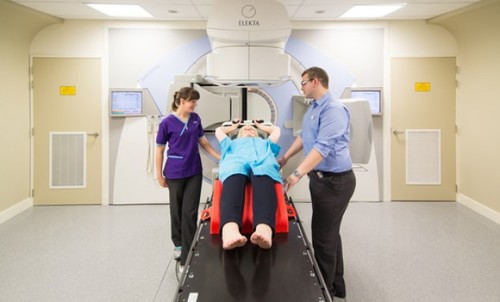
The most common way of delivering radiation to the breast is with a treatment machine called a linear accelerator, which delivers radiation beams from outside the body, targeting the whole breast or chest wall.
The patient is positioned on a bed and the head of the linear accelerator is lined up to focus the radiation to the targeted area. At each treatment appointment additional time is taken to ensure the correct positioning of the patient prior to treatment delivery. Once correctly positioned, the treatment takes only a few minutes to deliver. The head of the linear accelerator moves around the patient, delivering the beams. Patients do not feel the treatment being delivered.
Although the radiation therapists leave the room while the treatment is being given, they monitor the patients on closed circuit television and through microphones in the treatment room.
What to expect
Before going forward for treatment you will meet with a radiation oncologist, a cancer specialist who will plan and oversee your treatment. At this appointment, the benefits and risks of the treatment will be discussed with you so that you are able to make an informed decision about undergoing the treatment.
The next step is a planning or simulation appointment at the radiotherapy department. You will meet the staff involved in your care. This may include other doctors such as registrars and house surgeons, specialist oncology nurses, medical physicists, radiation therapists and support staff.
CT scans will be performed, with you in the position for treatment, to provide information about the structure of your breast and chest area so that an appropriate radiation plan can be calculated. This ensures that the most effective radiation dosage can be delivered to the targeted site while sparing unaffected tissues as much as possible.
To ensure that you are correctly positioned for treatment each day, at the time of CT simulation a variety of immobilisation devices may be used, such as lying on a headrest and breast board that will also be used during your treatment. In addition, small ink marks or tattoos will be placed on your skin to ensure that the radiation will be focused on the correct area each day (these marks are very small but can be removed at a later date if you prefer).
A typical course of radiation therapy is given daily (five days a week, excluding weekends, to allow the healthy cells to recover) for three to six weeks. The total radiation dose is divided into smaller daily doses, known as fractions, to reduce harm to healthy tissues.
Possible short-term side effects
Skin irritation in the treated area
This may range from mild “sunburn” to peeling or occasionally blistering. This side effect may not develop until treatment completion. It is usually at its most severe for one to two weeks and then settles over the following three to four weeks.
It’s important to try and reduce friction between your skin and clothing to reduce the risk of skin breakdown or blistering. You will be advised how to take care of your skin. For instance:
- Avoid hot water, lotions or other possible irritants on the skin in the treatment area. Using gentle soaps (non-perfumed, non-coloured) doesn't seem to increase skin irritation.
- Wear loose, light clothing (preferably non-synthetic materials such as cotton) over the area being treated.
- Avoid heat from hair dryers, electric hot pads, hot water bottles and sun in the treatment area.
- No adhesive tapes or sticking plasters should be applied to the skin in the treatment area
Some patients choose to use a transparent, breathable film dressing applied to the skin in the treatment area to try to reduce the skin reaction. Your radiation oncologist will be able to advise you on these products. Your radiation oncologist will also discuss with you the use of appropriate topical creams for the skin, depending on your skin reaction.
Breast heaviness, tenderness, itchiness, and swelling
This may occur towards the end of radiation therapy and continue for a few weeks after treatment completion.
Fatigue
Many people continue to work during their treatment but variable degrees of tiredness are common and may persist for a few weeks after treatment has finished. It’s important to plan your days so that you can have periods of rest if needed. Gentle exercise such as walking can be beneficial.
Possible long-term side effects
Changes to the look and feel of the breast
Radiation therapy may cause the breast to feel firmer and to be slightly smaller in volume. There may be some permanent discolouration of the skin in the treatment area and visible small red blood vessels (“spider veins”) may develop.
Your radiation oncologist will discuss with you the very small risks of long-term damage to normal tissue in the treatment area, such as changes in the lungs underlying the chest wall, rib fracture, heart problems if you have a left-sided breast cancer, nerve damage causing weakness in the arm, or lymphoedema of the arm or breast. There is an extremely small risk of inducing a second cancer in the treatment area many years after your breast cancer radiotherapy.
It is felt that the benefit of radiation therapy using modern radiation techniques outweighs the potential risks of treatment and your radiation oncologist will discuss this with you.
Heart disease
Fifty percent of breast cancers occur in the left breast and a small area of the heart can be exposed to the radiation field. Depending on the volume of heart treated and the dose delivered to the heart, this can increase the risk of heart disease over time.
Deep inspiration breath hold
Various techniques are used to minimise the exposure of the heart to radiation. One technique is to voluntarily hold a deep breath for 20-30 seconds while the radiation is delivered, as this expands the lungs and moves the heart away from the radiation field. Pre-treatment assessment of lung capacity and breathing patterns is carried out and the patient is given instruction on breath-holding for the required time.
To accurately maintain a deep inspiration breath hold, some centres use an active breathing co-ordinator device. Using this, patients are taught to take and hold a measured deep breath while the radiation dose is delivered. Using this method, patients can monitor their own breathing and the machine links to the linear accelerator ensuring that the radiation dose is only delivered when optimal breath hold is reached and maintained.
Both methods have been shown to reduce the exposure of the heart to radiation.
What's new in radiotherapy?
Dr. Maria Pearse’s radiation therapy presentation from 2016's Australasian Breast Congress Consumer Day.
Having radiotherapy for breast cancer
Watch this video made by Cancer Research UK.